Women's health and body require a careful and careful attitude. However, due to the fact that a modern woman lives in a frenzied mode, in which stresses and worries are constantly present, the female genital area suffers, first of all.
Modern medicine notes an increase in cases of diagnosing inflammation of the uterus. This is due to the fact that when symptoms are detected, a woman does not rush to see a gynecologist, referring to being busy. This approach is fundamentally wrong and entails irreversible consequences for the female body.
Today we will consider the main symptoms of uterine inflammation, discuss diagnostic, therapeutic and preventive measures.
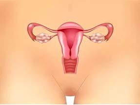
Female uterus
Before proceeding with the description of the disease, we will briefly consider the anatomical structure of a woman, more precisely, the uterus.
So, the female uterus means the muscular hollow organ that is located between the bladder and the rectum. The uterus acts as a receiving organ that accepts all the hormones produced by the ovaries. The surface of the organ is covered with a special layer, which undergoes significant changes at the moment of activation of the reproductive function. These transformations are caused by preparations for the fertilization process.
Infection routes
Due to the etiological factors described below, the mucous membrane of the organ can acquire an inflammatory nature. The development of the process may be associated with E. coli, chlamydia, or other pathogens.
In addition, pathogens are represented by viruses and protozoa. From the above it follows that endometritis occurs against the background of the penetration of infection into the organ. Below we will consider the methods of infection penetration.
So, infection occurs in the following ways:
- The first path is ascending.
- The second is lymphogenous.
- Hematogenous.
Often, the infection enters the organ through the external genitalia. After starting the infection process, the inflammatory process is activated. Due to the fact that the mucous membrane is tightly attached to the muscle layer, the process starts in the muscle tissue. In the event that inflammation is characterized by a chronic course, then the disease is identified as metroendometritis. The process has a short-term nature, which over time captures the uterine appendages or pelvic peritoneum.
Etiology of the disease
The endometrium includes the two layers described below:
- The first layer is functional.
- The second is basal.
The first layer peels off at the end of the menstrual cycle, while the second layer creates a new functional layer that starts the menstrual cycle again.

The main etiological factor is damage to the uterine lining. However, damage in itself does not pose a danger, viruses and virulent microorganisms that have entered the uterus are dangerous. Let's see what are the main reasons for the penetration of infection into the uterus.
So, inflammation develops for the following reasons:
- Improper douching.
- Complicated abortion and childbirth.
- Sexual intercourse during the menstrual cycle.
- Performing instrumental studies, in particular, laparoscopy.
We figured out the etiology, let's move on to the main symptoms of pathology.
Symptoms
As practice shows, the initial stages of the disease are characterized by the absence of symptoms. So that a woman can identify inflammation of the uterus, below is a list of the manifestations of the disease. It is worth noting that endometritis can be acute and chronic. An acute course is caused by mechanical damage, while a chronic one is the result of infection from a sexual partner.

Signs of acute uterine inflammation
Often, after seven days of diagnostic examination, childbirth, abortion, women notice the following symptoms:
- Bad condition.
- Refusal to eat.
- Headache.
- A significant increase in temperature, up to a febrile state.
- Aching and pulling pain in the lower abdomen.
- Atypical vaginal discharge.
- It is extremely rare, but uterine bleeding occurs.
If you find the above symptoms, you should immediately seek help from a gynecologist. There is one detail worth considering. Inflammatory pathologies tend to ectazate and involve adjacent organs in the process. The more a woman pulls to visit a doctor, the larger the scale of the lesion will turn out. Late and illiterate treatment is characterized by dire consequences.
Chronic symptoms
As a rule, a chronic course occurs against the background of late and illiterate treatment of the acute course of the pathology. As noted above, the chronic course has nothing to do with gynecological manipulations. The nature of this form of the disease is associated with sexual activity. Therefore, it is quite natural that chronic endometritis is characterized by those symptoms that give out the cause of the development of pathology.
The chronic course is represented by the following symptoms:
- Aching painful manifestations, localized in the lower abdomen.
- An increase in body temperature up to 38.5 degrees.
- Vaginal discharge. As for the nature of the discharge, it directly depends on the type of inflammatory process. Gonorrhea is accompanied by purulent discharge, trichomoniasis - copious and foamy discharge.
- Long menstrual cycle of the order of one week.
- Infertility and miscarriages.
Diagnosis
Diagnostic measures begin with taking anamnesis - determining the frequency and regularity of the menstrual cycle. Along with this, the doctor performs intrauterine intervention. Physical examination allows you to determine such abnormalities as an enlarged uterus, its induration, and so on.
Laboratory diagnostic methods are represented by blood tests and smears. Acute endometritis is characterized by an increase in the erythrocyte sedimentation rate and the number of leukocytes. In parallel with this, a C-reactive protein appears, indicating the presence of an inflammatory process in the body. Vaginal smear microscopy plays an important role in the diagnosis of the disease. Instrumental diagnostic methods are presented by ultrasound, biopsy and hysteroscopy.

Endometrial ultrasound
Ultrasound examination is fraught with some difficulties due to the characteristics of the anatomical structure of the upper part of the female genital organs. Despite some difficulties, this non-invasive research method is often used in diagnosing pathologies not only of the uterus, but also of the appendages.
By means of ultrasound, symptoms are determined only in 35 percent of cases. Inflammation of the uterus ultrasound often reveals indirect symptoms, however, they are all nonspecific and may indicate another disease.
On ultrasound, the following changes in the acute current are clearly visible:
- Quenching of the endometrium.
- Purulent clots.
The chronic form of the disease is characterized by pathological changes that are difficult to detect on ultrasound. However, this disease is accompanied by changes in the structure of the uterine mucosa, which are found on ultrasound.
For chronic endometrium, the following echoes are characteristic:
- Decrease and increase in the thickness of the uterine lining.
- Abnormal position of the uterus.
- Adhesions in the uterus.
Since inflammation can also affect the adjacent female genital organs, in particular, the ovaries and fallopian tubes, changes can be noted on ultrasound.
Despite this, ultrasound is an approximate diagnostic method, which, in combination with other methods, allows you to establish an accurate diagnosis. Ultrasound examination allows to exclude a number of diseases with identical symptoms. A complete and detailed examination includes ultrasound, biopsy, hysteroscopy.
Due to the fact that the diagnostic process includes an integrated approach, the data obtained during an ultrasound scan give the right to make a diagnosis such as endometritis.
Healing activities
Endometritis treatment begins immediately after an accurate diagnosis is made. In the event that the pathology was detected in a timely manner, then the treatment takes place on an outpatient basis, but under the obligatory supervision of the attending physician. Otherwise, the patient is referred to the hospital. As for the treatment regimen, in most cases it is the same and consists of the following points:
- Antibacterial therapy.
- Mechanical cleaning of the uterine cavity.
- Detoxification.

How is acute endometritis treated?
If the gynecologist detected an acute process in time, then the treatment avoids the development of dangerous complications, including problems with pregnancy. Fortunately for women, this disease does not belong to the list of incurable diseases. The treatment takes place in several stages.
At the first stage, a woman is prescribed immunomodulators and a complex of vitamins. At the second stage, the attending physician prescribes a course of antibiotic treatment. Antibiotics are given both intravenously and intramuscularly. As a rule, the course of treatment is about 5 days. In fact, the duration of treatment depends on the stage of the disease.
To relieve severe pain, patients are prescribed pain medications. Together with antibiotics, therapy includes the appointment of anti-inflammatory drugs, in particular, Aspirin, Ibuprofen. The third stage involves cleaning after abortion, childbirth, and cesarean section. The final stage is physiotherapy.
Chronic course: therapy
As noted above, the chronic stage is characterized by a different nature of occurrence, therefore, the goal of therapy is to eliminate a specific causative agent of the disease. To find a suitable and effective treatment regimen, specialists resort to taking a smear for culture. This allows you to accurately determine the sensitivity of the pathogen to various medications. Based on the results of such a study, a list of effective drugs is formed. Therapeutic measures for the chronic course of the pathology are based on antibacterial and antiviral treatment.
Important! One of the most effective methods of treatment is considered to be the introduction of a drug into the lining of the uterus. This approach allows you to influence the focus of inflammation.
So, therapy is based on the following manipulations:
- On hormonal treatment. Oral contraceptives are prescribed for women.
- On splitting adhesions through surgery.
- Physiotherapy. Physiotherapy plays an important role in the treatment of endometritis. It allows not only to enhance the outflow of mucus, pus from the uterine cavity, but also to normalize the work of the ovaries.
As for the purulent form of the disease, the therapy is based on mechanical cleaning of dead tissue. Of course, this procedure is unpleasant, so the woman is given general anesthesia.
Spa assistance
Of course, any disease requires the restoration of the body. Moreover, when it comes to the female body. Spa therapy combines healing and restorative measures. However, such treatment can only be started during remission.
Complications
The inflammatory processes occurring in the female body worsen the standard of living. Through strong painful sensations that gain intensity during intercourse, a woman's sex life suffers. In parallel with this, the infection through the lymphatic and circulatory system can spread throughout the body.
One of the most dangerous complications of the disease is sepsis. As you know, the neglected form is accompanied by the formation of adhesions in the small pelvis. Adhesions in the body lead to a prolonged lack of conception of a child or to problems with bearing a fetus. Violated uterine the process is accompanied by a violation of the menstrual cycle. Therefore, in order to avoid the above complications, it is recommended to adhere to simple preventive rules.
Preventive actions
In fact, prevention is based on simple and straightforward rules. So, prevention includes the following activities:
- Lead a healthy lifestyle.
- Strengthen the immune system.
- Use condoms during intercourse. Today they are not only reliable, but also absolutely safe contraceptives.
- Adhere to the rules of personal hygiene.
- Systematically undergo examinations by a gynecologist.
- Have sex with only one partner.
Note that any disease is easier to prevent than to cure. When the first symptoms of the disease appear, postpone all cases, no matter how important they were, and visit a gynecologist. After all, the purpose of any woman is to become the mother of her beloved baby. Good health and happy healthy minutes.
Identification of appendicitis is possible in different ways of diagnosis. In cases where the symptomatology is poorly expressed, a latent disease, the inflammatory process can be determined using an ultrasound examination. The reliability of the diagnosis is over 90% and allows you to detect the presence of problems associated with other organs of the patient. Is it possible to see appendicitis on ultrasound, the advantages and disadvantages of diagnosis, the features of the procedure in women and children, are discussed in the article.
The manifestations of inflammation of the appendix in most cases have pronounced symptoms. The general picture of the patient's condition, complaints of pain and the location of the focus of pain clearly indicate the diagnosis. The development of the disease proceeds rapidly, the attack turns into complications in the form of peritonitis, sepsis, gangrene. These options require urgent care and surgery. But there is a sluggish course of inflammation, in which the patient does not notice the usual symptoms. These cases make wave diagnostics an irreplaceable research method.
Symptoms similar to acute appendicitis occur with inflammation of other abdominal organs. It is important to differentiate the conditions to determine the true diagnosis of the patient. Often, the situation is complicated by the atypical location of the appendix of the cecum in the abdominal cavity. With an atypical location, it is difficult to determine the location of the pain focus. In these cases, ultrasound of appendicitis determines the correct diagnosis and allows you to prescribe an adequate course of treatment.
Advantages:
- Security. Unlike diagnostics using X-ray irradiation, diagnostics using an apparatus with an ultrasonic sensor does not harm health. This is especially true when examining pregnant women and children.
- Availability. Many experts are inclined to believe that CT has the most reliable diagnosis. However, the results of ultrasound research are no less accurate, but at the same time more affordable and cheaper.
- Speed. In case of an emergency, it is necessary to save as much time as possible. The procedure does not require additional preparation before carrying out. Ultrasound shows the patient's condition in real time.
The relevance of the use of ultrasound in diagnostics is high for pediatric patients and for women in pregnancy. Children, due to their age and lack of experience, cannot describe the exact picture of pain and the location of uncomfortable sensations. Pregnant women at risk due to displacement of the appendix to an atypical position by the increased volume of the uterus. Both cases involve examination with ultrasound waves, which in these cases is called the most accurate diagnostic method.
With all the advantages, there are a number of disadvantages. By ultrasound, it is impossible to consider the focus of inflammation if the patient is overweight. A large accumulation of gases in the intestine also makes it difficult to diagnose due to the inability to consider the problem area.
What does ultrasound show?
An ultrasound scan does not involve preparation. The procedure is performed using a special sensor through the peritoneal wall. Sometimes women undergo a vaginal examination with an atypical location of the organ or with gynecological problems in the form of an inflammatory process in the ovaries.
During the study, the doctor discovers the cecum, from which the appendix branches off, even with its atypical location. When examining the appendix, the doctor can detect the presence of an inflammatory process in the appendix of the cecum and adjacent organs of life. A number of factors indicate the inflammatory process of the appendix:
- Uneven thickness of the walls of the organ.
- Increase in size.
- The presence of fluid in the branch of the cecum.
- Omentum inflammation.
- Abscesses of the peritoneum.
- Layering and discontinuity of the appendix structure.
An ultrasound diagnosis is prescribed in the presence of constant aching pains, nonspecific for inflammation of the appendix. For differentiating acute appendicitis from diseases of other organs of the patient, especially from gynecological problems, this study is irreplaceable. It shows especially reliably atypical symptoms in chronic inflammation.
Procedure
With a weak severity of pain, with an atypical location of the painful area, the analysis is carried out in the following way:
- Study of the apex of the cecum.
- Detection of the iliac vessels.
- Study of the acne muscle.
- Study of the place behind the cecum.
- Analysis of the state of the peritoneal and pelvic organs.
- The right ovary is examined especially for women.
The final diagnosis is made by the doctor. There is not always enough data to make an adequate diagnosis. Some cases require additional tests, diagnostics using MRI, laparoscopy, or CT. Conclusions are made based on the results of all types of research.
Often, ultrasound is prescribed after removal of the appendix, especially if complications arise or a sharp deterioration in the patient's condition, allowing you to see the internal source of the complication.
Carrying out in women
The female body is arranged somewhat differently. Therefore, the appearance of pain characteristic of appendicitis may indicate gynecological problems in the form of inflammation of the appendages or ectopic pregnancy. Therefore, the doctor cannot make the correct diagnosis only on the basis of probing the peritoneum and the results of blood and urine tests. This is exactly the option where ultrasound is irreplaceable.
A clear source of pain is seen during the procedure. Examination of the peritoneal and pelvic organs displays the condition of the appendages and appendix, allowing you to diagnose the exact cause of the ailment. The procedure for examining female patients is carried out more often due to the structural features of the female body. The internal reproductive structure of women, the organs of the urinary system are in close contact with the digestive system. As a result, inflammatory processes in the gynecological parts of the woman's body are transferred to the genitourinary system or intestines.
In addition, during menstruation, a woman's uterus swells, increases in volume and displaces other organs in the abdominal cavity. During pregnancy, the uterus increases many times, in addition to displacement, it squeezes and disrupts blood circulation in the gastrointestinal tract, which leads to inflammatory processes. In this case, ultrasound remains a relevant research method capable of showing an accurate picture.
Features of the procedure in childhood
Children are not always able to describe the symptoms of an attack, they cannot indicate where the pain is located. An ultrasound examination in children is a safe and quick way to establish the cause of pain and an accurate diagnosis. The attack itself develops in a child much faster than in adults.
This is due to the physiology of the structure of the child, the course of metabolic processes. An attack may be triggered by ARVI or tonsillitis, malnutrition, dysbiosis, gastritis. Ultrasound will show the focus of inflammation and the cause of its development.
In some cases, the removal operation is contraindicated, antibiotic therapy is prescribed. Gradually appendicitis becomes chronic. The described moment involves a mandatory ultrasound examination two or three times a year to monitor the condition of the appendix. This is necessary because the disease in a chronic condition can proceed secretly and lead to aggravating consequences for human health.
Ultrasound diagnostics is an informative, safe, affordable method for detecting pathology in acute appendicitis. The procedure will help the doctor to differentiate malaise from other diseases, see inflamed processes, exclude or determine the presence of problems in the adjacent organs of the abdominal cavity. Diagnostics can make it possible to establish the correct diagnosis, and in case of insufficient data, it is supplemented by laboratory tests, MRI or CT.
It is important to take good care of your health, monitor your physical fitness and well-being, and seek help in time for ailments. Strong immunity will allow you to maintain health, prevent the development of pathological conditions and complications, and ensure a speedy recovery from diseases. Well, if the body malfunctioned and medical assistance was required, modern research methods in the form of diagnostics using ultrasound will help to make an accurate diagnosis, to identify the true cause of the disease.
Ovarian inflammation ( oophoritis) is an acute or chronic pathological process that affects the tissue of the female reproductive glands, causing a disorder of their function. In the overwhelming majority of cases, this ailment does not develop independently, but in combination with the inflammatory process within the fallopian tubes ( so-called adnexitis). In foreign literature, acute inflammation of the ovaries is usually combined with inflammation of the fallopian tubes ( salpingitis) into one common clinical syndrome - inflammation in the small pelvis.
In most cases, inflammation of the ovaries and uterine appendages occurs due to the penetration of various infectious agents, more often - the causative agents of sexually transmitted diseases. For this reason, oophoritis and salpingitis usually develop in young women under the age of 25, who are sexually active and do not use barrier methods of contraception ( condoms).
Inflammation of the ovaries, provoked by pathogenic bacteria or viruses, is a dangerous and serious ailment that causes reproductive disorders ( infertility), as well as hormonal disruptions due to changes in endocrine activity ovaries... With an aggressive course of the disease, local or widespread purulent complications may occur that pose an immediate threat to a woman's life.
Interesting Facts
- inflammation of the ovaries and fallopian tubes is one of the most common causes of female infertility;
- inflammation of the ovaries quite often occurs against the background of the asymptomatic course of certain sexually transmitted diseases ( chlamydia);
- inflammatory process in the pelvic area is more common among young women;
- isolated damage to the ovaries by an infectious or inflammatory process practically does not occur;
- ovarian inflammation can occur in response to an inflammatory process in other organs;
- hormonal disruptions increase the likelihood of penetration of infectious agents into the upper parts of the female reproductive system;
- stress is a factor that significantly weakens the protective potential of the female body and increases the risk of damage to the fallopian tubes and ovaries.
Anatomy of the uterine appendages
 The female genital organs are conventionally divided into upper and lower sections. This division simplifies the systematization of the clinical manifestations of some genital infections, and also allows a better understanding of the mechanisms of penetration of pathogenic agents.
 The lower sections of the female genital organs are represented:
- Vulva. The vulva is the labia majora and labia majora, which act as the entrance to the vagina, clitoris, and the mouth of the urethra.
- Vagina.The vagina is a tubular muscular-elastic organ that performs sexual, reproductive, protective and excretory functions. Normally, the vaginal environment is not sterile and is formed by Doderlein bacilli and a number of other saprophytic ( non-pathogenic) microorganisms. Due to this, the vagina has its own microflora, which contributes to its protection in the event of any infections entering and developing.
- Cervix.The cervix is \u200b\u200bthe portion of the uterus located between the vagina and the uterine cavity. A canal passes through the cervix, which is normally closed and contains cervical mucus, which protects the overlying structures from infection.
- The ovaries.The ovaries are paired female sex glands that are located in the pelvic cavity and perform reproductive and hormonal functions. Produce sex steroid hormones ( estrogen and progesterone). The process of maturation of the egg takes place in the ovaries.
- Uterus.The uterus is a muscular organ located in the pelvic cavity. Performs reproductive function ( carrying a pregnancy) and menstrual ( detachment of the inner mucous membrane). Through the fallopian tubes, the uterus connects to the abdominal cavity, and through the cervical canal - to the vagina and the external environment.
- Uterine ( fallopian) pipes.The fallopian tubes are a paired organ that is located in the pelvic cavity and connects the uterus to the abdominal cavity. In the lumen of the fallopian tubes, fertilization of the egg takes place, and their main function is to transport the embryo or egg into the uterine cavity.
The ovaries are supplied with blood by the ovarian artery, which originates from the abdominal aorta, and also by the branches of the uterine artery. Venous blood flows through the ovarian vein, which forms the ovarian plexus, into which blood also flows from the fallopian tubes. Knowledge of the characteristics of the blood supply allows a better understanding of the possible mechanisms of penetration of infectious agents to the ovaries.
The ovaries are innervated by the branches of nerves from the lower hypogastric plexus. The ovaries are not covered with the peritoneum, but they are in close contact with it. These facts are of great importance for understanding the mechanisms of the onset of pain during the development of the inflammatory process.
Next to the ovaries is the bladder, bowel loops, appendix, rectum. These formations may not be directly adjacent to the ovaries, but in some conditions they can serve as the initial source of infection or inflammation.
Ovarian inflammation causes
 The most common cause of inflammation in the ovaries is infection. However, this is far from the only reason that can provoke this ailment. The inflammatory process is a defense mechanism that arises in response to the action of any damaging factor and is aimed at reducing damage. Based on this, it can be assumed that the inflammatory response can occur in response to many pathological situations.
 Ovarian inflammation can occur in the following situations:
- Infections.In the overwhelming majority of cases, the inflammatory process in the ovaries occurs due to the penetration of an infection, which can be of a bacterial, viral or fungal nature. Most often, oophoritis is associated with sexually transmitted diseases, but it can also occur with tuberculosis and with some nonspecific infectious processes. It should be understood that the infection rarely covers only the ovaries and usually affects either the uterus, or the fallopian tubes, or both organs at the same time and only then covers the ovaries. However, in some cases, the infection can penetrate into the ovaries and from other organs by direct contact with an infectious-inflammatory focus or by the introduction of pathogenic agents along with the blood stream.
- Mechanical damage.Trauma to the ovaries, fallopian tubes or uterus can cause an inflammatory process that can cover the ovaries, as well as significantly weaken local immunity and become a factor predisposing to infection.
- Inflammation of adjacent organs.The ingress of biologically active pro-inflammatory substances into the ovaries can provoke some inflammatory reaction.
- Necrosis and inflammation of neoplasms ( tumors). With the development of some tumors, a necrotic process can occur, which can trigger an inflammatory reaction.
Sexually transmitted infections
In most cases, the inflammatory process in the pelvic cavity, covering the fallopian tubes and ovaries, is associated with sexually transmitted infections. Most often, the disease is associated with bacterial lesions caused by pathogens of gonorrhea or chlamydia, but other pathogenic agents may also occur.Oophoritis can be caused by the following pathogens:
- Gonococci.Gonococci are the causative agents of gonorrhea, one of the most common sexually transmitted diseases. These microorganisms enter the reproductive system during unprotected sex with an infected partner. Initially, they affect the lower parts of the genital tract, however, with a weakening of local or general immunity, as well as with the development of a number of predisposing factors, they can penetrate into the uterine cavity, go to the fallopian tubes and cause infection of the ovaries.
- Chlamydia.Chlamydia is the causative agent of chlamydia, a common venereal disease characterized by a latent course. Like gonorrhea, this ailment is transmitted during unprotected sex, but unlike it, chlamydia rarely causes any severely disturbing symptoms. For this reason, this infection is often diagnosed already at the stage of development of various complications, including oophoritis.
- Trichomonas.Vaginal Trichomonas is the causative agent of trichomoniasis, a genital infection that, according to the World Health Organization, is the most common among humans. As well as chlamydia, trichomoniasis of the lower genital tract is quite often asymptomatic or with minor clinical manifestations. This creates the prerequisites for the spread of the infectious process into the uterine cavity and its appendages. Trichomoniasis rarely affects the ovaries, but the damage to the fallopian tubes caused by it can, in one way or another, cause an inflammatory reaction in the female genital glands with a violation of their function.
- Mycoplasma.Mycoplasmas are small bacteria that can cause mycoplasmosis. These microorganisms are conditionally pathogenic, in other words, they can cause a disease only with a significant violation of the general condition of a woman and with a decrease in her local or general immunity. They are transmitted during sexual intercourse, as well as in some types of household contacts. Mycoplasmosis is characterized by a chronic malosymptomatic course. Penetration into the upper parts of the reproductive system is accompanied by the appearance of signs of severe damage to the genital organs.
Initially, the infectious process affects the external genital organs ( small and large labia and adjacent glands), as well as the urethra and vagina. It should be noted that normally the vagina is inhabited by Doderlein bacilli, which form its normal environment and perform a protective function, since they do not allow pathogenic microorganisms to colonize this organ. However, in some situations, the vaginal microflora and its protective potential can be violated, which creates the prerequisites for the development of infection.
Risk factors for infection of the lower genital organs are:
- incorrect intake of antibiotics;
- douching the vagina;
- lack of personal hygiene;
- stress;
- diseases of the immune system;
- frequent change of sexual partners;
- unprotected sex.
The risk factors for the spread of infection to the upper sections of the female reproductive system are:
- abortion;
- therapeutic or diagnostic curettage of the uterus;
- installation of intrauterine contraceptives ( spirals);
- spontaneous abortion;
All these factors are due to the fact that the expansion of the cervical canal and the removal of the mucous plug opens the way for infectious agents located in the vaginal cavity.
In the future, the infectious process covers the lining of the uterus, and then the fallopian tubes and ovaries. In some conditions, pathogenic agents can cause the formation of purulent infectious and inflammatory foci in the uterine appendages, which is fraught with a serious violation of the general condition and is associated with a high risk of developing systemic complications.
Additionally, the hematogenous route of penetration of infectious agents into the ovaries is considered. This is due to the peculiarities of the blood supply to the ovaries, which receive part of the arterial blood from the branches of the uterine artery. Due to this, pathogens that can be in the blood of a person for a short or long time can be brought into the ovaries along with the bloodstream from the lower parts of the reproductive system.
Viral lesion
It is assumed that the inflammatory process in the ovaries can be triggered not only by bacteria, but also by viruses. There are a number of studies that indicate that at least two sexually transmitted viral infections can provoke inflammation in the uterine appendages.Ovarian inflammation can be caused by the following pathogens:
- Herpes simplex virus type 2... Herpes simplex virus type II, also known as genital herpes, can enter the body through defects in the skin, as well as through the mucous membranes of the genitals during unprotected sexual contact with an infected person. It has the ability to integrate into human cells, due to which a complete cure becomes impossible. Because of the scanty symptoms, genital herpes is a fairly common infection. During the activation period, the virus causes local foci of necrosis of the mucous membrane, which provokes an acute inflammatory reaction.
- Cytomegalovirus. Cytomegalovirus comes from the same family as herpes simplex virus. It is capable of affecting many organs, including the genitourinary system. In most cases, it does not pose a particular danger, however, against the background of reduced immunity, it can cause serious complications. It can cause inflammation of the pelvic organs, both alone and in combination with a number of other pathogens ( usually bacterial).
Separately, it is necessary to mention the human immunodeficiency virus ( HIV), which does not independently affect the upper sections of the female reproductive system, but due to its ability to weaken the immune system creates the prerequisites for infection with other pathogens. In addition, against the background of HIV infection, especially at the stage of acquired immunodeficiency syndrome ( AIDS), optimal conditions are created for the infection of the genitals, including the ovaries, not only with aggressive pathogenic microorganisms, but also with opportunistic pathogens, which are normally harmless to humans.
Tuberculous lesion
Tuberculosis is a common infectious disease caused by Mycobacterium tuberculosis. In most cases, this disease affects the lungs, but in some cases, the formation of foci in other organs is possible.Typically, TB infection occurs by inhalation of sputum particles containing a tubercle bacillus ( airborne transmission), however, it is possible for the pathogen to penetrate when eating contaminated food ( milk and dairy products), as well as through the skin ( rarely). In conditions of reduced immunity or impaired body resistance, the tubercle bacillus begins to multiply and develop in the lung tissues, provoking a specific inflammatory reaction. As a result, a primary complex is formed, from which pathogens, along with the blood flow, can enter the bones, kidneys, eyes, skin and genitals.
The penetration of mycobacterium tuberculosis into the genitals is due to the peculiarities of their blood supply. Since the fallopian tubes and ovaries receive blood from the branches of the uterine and ovarian arteries, at their intersection ( so-called anastomoses) the speed of blood flow slows down, and this creates ideal conditions for the penetration of bacteria into these organs. The hematogenous path of spread is associated with a predominantly bilateral lesion of the uterine appendages.
Infection with tuberculosis sexually is considered impossible, since the vaginal environment is extremely unfavorable for mycobacterium tuberculosis. However, if the pathogen enters the injured or inflamed mucous membranes of the lower parts of the reproductive system, primary infection of the genitals can occur.
The main problem of tuberculous lesions of the fallopian tubes and ovaries is that this disease in the vast majority of cases is asymptomatic. Women rarely seek medical help for this infection. This leads to the fact that, against the background of a long course of the disease, various complications and irreparable structural and functional damage develop.
Mechanical damage to the uterine mucosa and fallopian tubes
The inflammatory process, as mentioned above, is a kind of protective reaction of the body, which is aimed at reducing the damaging effect of any traumatic factor. Thus, not only a bacterial or viral infection, but also mechanical damage can trigger an inflammatory reaction in the uterine appendages.Mechanical damage to the ovaries and fallopian tubes is possible in the following situations:
- Blows to the abdomen.Exposure to a short but strong impulse can cause contusion of many internal organs, including the uterus, fallopian tubes and ovaries. Under the influence of a damaging factor, local structural damage can occur, partial or complete destruction of blood vessels with impaired local blood circulation is possible. To minimize the effects, the body triggers an inflammatory response, which in some cases can cause even more severe damage.
- Penetrating wounds in the abdomen.Penetrating wounds to the abdomen can damage the upper sections of the female genital organs, which can lead to inflammation. In addition, most penetrating wounds are potentially infectious.
- Surgical interventions on the abdominal and pelvic organs.Any surgical intervention, no matter how minimally invasive it may be, traumatizes the internal organs to one degree or another. Strong pressure on the genitals through surgical instruments, cutting them or burning them down can provoke an inflammatory reaction. In addition, do not forget about foreign materials that may be in the area of \u200b\u200bthe operation ( suture material, various prostheses, stents, gases and solutions) and also cause inflammation.
- Invasive gynecological procedures.Gynecological procedures that involve instrumental effects on the internal genital organs ( abortion, curettage) are associated with some injuries, which directly provoke an inflammatory response. In addition, they reduce local immunity and create prerequisites for the penetration of infectious agents.
Inflammation of adjacent organs
The defeat of the ovaries may be associated with an inflammatory process that has engulfed neighboring organs. Most often this is caused by the transfer of bacteria from the primary infectious focus through the organ wall, but it can also occur for a number of other reasons.The ovaries can be involved in the inflammatory process when the following organs are damaged:
- Colon.Inflammation of the large intestine, known as colitis, is usually caused by an imbalance between normal and pathogenic intestinal microflora ( pathogenic bacteria begin to dominate). In some cases, the intestinal wall can be depleted, ulcers and even through holes can form in it ( which leads to the development of peritonitis and is extremely dangerous). In addition, inflammation in the intestines is accompanied by edema, slowing blood flow and dysfunction. Under the influence of these factors, there is a risk of pathogens passing through the intestinal wall to neighboring organs - the peritoneum, ovaries and fallopian tubes, other parts of the intestine.
- Appendix.Inflammation of the appendix ( appendicitis) is one of the most common surgical pathologies. There are several theories explaining the mechanism of development of this disease, but regardless of the initial cause, the developing inflammatory reaction affects the entire thickness of the muscular wall of the organ and covers part of the serous membrane covering it. The resulting pathological reaction is quite massive, and in contact with other organs can cover them.
- Bladder.Bladder infection ( cystitis) in some cases can cause an inflammatory process in the ovaries. However, in the vast majority of cases, the ovaries are involved in the inflammatory process not because of contact with the bladder, but because of the parallel lesion of the internal genital organs and the bladder by sexually transmitted infections.
- Peritoneum.The peritoneum is the serous membrane that covers most of the abdominal organs and lines the walls of the abdominal cavity itself. Despite the fact that the ovaries are not covered by the peritoneum, an infectious and inflammatory process on the surface of the peritoneum can cause damage to the ovaries. However, much more often everything happens the other way around, and inflammation of the ovaries causes local inflammation of the peritoneum - pelvioperitonitis. It should be understood that peritonitis ( inflammation of the peritoneum) is an extremely serious condition that requires immediate medical treatment.
Ovarian inflammation symptoms
 The clinical manifestations of ovarian inflammation are quite diverse, but they are nonspecific, as they are similar to the symptoms of diseases of other pelvic organs.
 The symptoms of ovarian inflammation are formed by the inflammatory reaction itself, which, one way or another, changes the function and structure of the organ, as well as infectious agents, which in most cases are the cause of oophoritis.
Ovarian inflammation is accompanied by the following symptoms:
- lower abdominal pain;
- increased body temperature;
- disruption of the gastrointestinal tract;
- violation of the menstrual cycle;
- painful sexual intercourse;
- hormonal disorders;
- infertility;
- pain in the upper abdomen;
- muscle tension of the anterior abdominal wall.
Lower abdominal pain
Lower abdominal pain is the main symptom of acute inflammation of the ovaries and fallopian tubes. Pain occurs due to a slight increase in organ size due to edema, as well as due to the effect of pro-inflammatory biologically active substances on sensitive nerve endings. Since the ovaries are innervated by the branches of the hypogastric plexus, the resulting painful sensation is usually a pulling, aching character. When involved in the inflammatory process, visceral ( covering organs) of the peritoneum, the intensity of pain increases slightly, and reflex vomiting may occur. If the infectious-inflammatory focus covers the parietal ( parietal) the peritoneum, the pain increases significantly, becomes sharp, reflex muscle tension occurs.The duration of the pain varies depending on the activity of the inflammation and on the treatment taken. Pain is usually present for at least 2 to 3 days, but not more than 3 to 4 weeks.
Increased body temperature
An increase in body temperature is a nonspecific reaction of the body that occurs in response to the penetration of any foreign protein. Fever is aimed at creating conditions that are unfavorable for the pathogenic agent, but optimal for the functioning of the immune system. The body temperature rises as a result of the effect of a number of biologically active substances formed in the focus of inflammation on the structures of the central nervous system. Fragments of pathogens, particles of foreign proteins, as well as pyrogenic ( substances that can increase body temperature), formed during immune reactions.There are three stages in the development of fever:
- The rise in temperature.The rate of temperature rise depends on the nature and properties of the pathogen. With a sharp rise, a feeling of chills arises, which indicates the activation of heat-saving mechanisms ( reduced sweating, "goose bumps", peripheral vascular contraction). Body temperature rises due to increased thermogenesis ( muscle tremors, accelerated metabolism of nutrients).
- Plateau stage.At the plateau stage ( keeping body temperature) the feeling of chills disappears and the body temperature stabilizes. Depending on the pathogen, the body temperature with inflammation of the ovaries can rise to 37.5 - 38 or even up to 39 degrees. With the development of complications, the body temperature can exceed 39 degrees.
- Decrease in temperature.A decrease in body temperature can occur either gradually or abruptly. The body temperature decreases after the elimination of the action of pyrogenic substances, when taking certain medications, as well as when the body is severely depleted.
Disruption of the gastrointestinal tract
Inflammation of the ovaries or other parts of the upper genital tract can cause various disorders of the gastrointestinal tract.The following disorders of the gastrointestinal tract may occur:
- Nausea and vomiting. Nausea and vomiting occur reflexively, in response to intense painful stimulation of the hypogastric plexus. In addition, nausea is one of the possible consequences of an increase in temperature and general intoxication of the body. Vomiting is usually mild, not associated with food intake. Abundant vomiting that does not bring relief indicates the possible development of complications ( peritonitis).
- Diarrhea. Diarrhea occurs due to intoxication of the body, as well as due to irritation of the intestines with an inflammatory focus.
- Desire to defecate. Frequent urge to defecate occurs due to irritation of the ampullar part of the rectum with an inflammatory focus in the genitals and in the area of \u200b\u200bthe peritoneum located in the small pelvis.
Pathological discharge from the genital tract
Normally, discharge from the genital tract is a small amount of odorless transparent or whitish mucus, the release of which is not accompanied by any unpleasant sensations.In the presence of infectious and inflammatory foci within the upper or lower genitals, various pathological vaginal discharge often occurs, indicating an ailment. The nature of the discharge depends on the nature and properties of the pathogen, as well as on the localization of the lesion and the body's resistance.
It should be understood that discharge can form in the vagina, cervix, and uterine cavity. An infectious and inflammatory process limited to the fallopian tubes or ovaries is extremely rarely accompanied by discharge from the genital tract, since more often in this case, pathological fluids are drained into the pelvic cavity.
The following options for pathological discharge from the genital tract are possible:
- Purulent discharge. Purulent discharge is a specific symptom indicating the bacterial nature of pathogens. They are a yellowish-green viscous liquid, the amount of which can vary depending on the severity of the process and the aggressiveness of the pathogen. Purulent discharge is characterized by an unpleasant smell of rotten fish. When anaerobic microflora is attached, purulent discharge becomes foamy, since these microorganisms produce gas, which foams the pus.
- Serous discharge. Serous discharge is characteristic of viral lesions of the cervix and uterus. They arise due to vasodilation and the release of part of the plasma from the bloodstream during an inflammatory reaction. Usually, such discharge is transparent or slightly yellowish, odorless.
- Bloody issues. Bloody discharge occurs when the vessels are melted by pathogenic agents or when their integrity is destroyed during an inflammatory reaction. Bloody discharge is usually scanty, not abundant, represented by dark blood, occurs regardless of the menstrual period. Discharge may be accompanied by pain in the lower abdomen.
Disruption of the menstrual cycle
The menstrual cycle is a periodic change in a woman's genitals, aimed at maintaining readiness for conception. This process is regulated by hormones of the ovaries, hypothalamus and pituitary gland.At the heart of the menstrual cycle is the periodic renewal of the uterine lining and the maturation of the egg. This happens in several phases, each of which is regulated by certain hormones. First, there is a detachment of the uterine mucosa ( endometrium), which is accompanied by bleeding. Subsequently, under the action of sex hormones, the regeneration of the mucous layer begins in the uterine cavity, and a dominant follicle is formed in the ovaries. Subsequently, by the time of ovulation, when the follicle ruptures and the level of estrogen and progesterone rises, the uterine mucosa thickens significantly, and the egg released from the follicle ( which at this stage it is more correct to call the first-order oocyte) migrates through the fallopian tubes into the uterine cavity. If fertilization does not occur during this period, then this cycle is repeated again.
With inflammation of the ovaries, the menstrual cycle can be disrupted for the following reasons:
- damage to the lining of the uterus;
- a decrease in the level of sex hormones due to dysfunction of the ovaries;
- ovulation disorders;
- violation of endometrial regeneration.
- lack of discharge during menstruation;
- scanty discharge during menstruation;
- profuse discharge during menstruation;
- long menstrual cycle;
- pain during menstruation.
Painful intercourse
The inflammatory process in the pelvic cavity is often accompanied by pain during intercourse. This is usually associated with damage to the vagina, but it can also occur with damage to the upper reproductive system.Soreness during intercourse is associated with excessive vaginal dryness, which occurs either due to an inflammatory lesion of the vagina itself, or due to a decrease in the level of the sex hormone estrogen. As a result, due to insufficient moisture, friction increases and a painful sensation arises during sex. This leads to the fact that the woman's sexual desire decreases ( decreased libido), mood is disturbed, depression may develop.
Hormonal disorders
Hormonal disorders during inflammation of the ovaries are not always found, but in some situations they are quite possible. They arise due to structural and functional rearrangements of the organ, which leads to a decrease in the synthesis of sex hormones ( estrogen and progesterone).Since one normally functioning ovary is able to maintain the level of sex hormones within the physiological norm, hormonal disorders occur only with bilateral organ damage or with damage to the only working ovary.
Hormones are known to regulate many physiological processes in the human body. With a decrease in the level of sex hormones, a disorder of sexual and reproductive function occurs, as well as disorders of the central nervous system ( mood changes, depression, manic-depressive states), of cardio-vascular system ( heart rhythm disturbances, high blood pressure) and from the side of metabolism ( obesity occurs, cholesterol levels rise). Of course, some of these manifestations can develop only in the case of a protracted course of the inflammatory process, accompanied by hormonal disruption.
Infertility
Infertility is one of the most frequent consequences of the inflammatory process in the area of \u200b\u200bthe uterine appendages and often acts as the main reason for a woman to seek medical help.Infertility with damage to the ovaries is associated with impaired production of eggs, as well as developing hormonal disorders. However, much more often infertility occurs due to damage to the fallopian tubes, which in the vast majority of cases accompanies oophoritis. Due to the inflammatory reaction, the fallopian tubes are narrowed, functional and structural changes occur in them, which lead to partial or complete obstruction for the egg and sperm.
Muscle tension of the anterior abdominal wall
The tension of the muscles of the anterior abdominal wall occurs when the parietal is involved in the inflammatory process ( parietal) the peritoneum. Muscle contraction occurs reflexively, in response to strong painful stimulation emanating from the inflammatory focus. Due to muscle tension, tension and irritation of the peritoneum decreases, which makes it possible to somewhat alleviate the pain sensation.In addition to the symptoms listed above, the inflammatory process with localization in the ovaries and fallopian tubes can be accompanied by a number of other signs, which in most cases arise already at the stage of development of complications.
Inflammation of the uterine appendages may be accompanied by the following signs of a complicated course:
- Pain in the upper abdomen and in the right hypochondrium.A painful sensation in the right hypochondrium, which arose against the background of pain in the lower abdomen, temperature and other signs of damage to the female reproductive system, indicates the occurrence of perihepatitis - inflammation of the liver capsule ( fitz-Hugh-Curtis syndrome). It is characterized by some dysfunction of the liver, an increase in the level of liver enzymes, and sometimes by yellowness of the skin and mucous membranes.
- Swelling of the abdomen from the side of the lesion.The appearance of abdominal swelling on the side of the affected ovary, which can be determined visually or during palpation, indicates the development of a tubo-ovarian abscess, a cavity filled with purulent contents. It is a potentially dangerous condition that requires surgical treatment.
Diagnosis of ovarian inflammation
 Diagnosis of ovarian inflammation is a difficult task due to the fact that this disease has symptoms similar to some other ailments, and also due to the fact that the inflammatory reaction is extremely rarely limited to the ovaries alone, involving the fallopian tubes, uterus and other parts of the genital systems. This creates additional difficulties in diagnosing the disease.
 Before starting any diagnostic procedures, a conversation with a doctor is held, during which the main symptoms, the time of their onset, intensity, and main characteristics are clarified. Data is collected on previous surgical operations, on known acute and chronic diseases. The doctor finds out whether the menstrual cycle is regular, when were the last menstruation, what is the amount of discharge during menstruation, whether the menstrual period is accompanied by pain or discomfort.
An inflammatory process in the uterine appendages is suspected in the presence of the following signs:
- lower abdominal pain;
- pathological discharge from the genital tract;
- increased body temperature;
- hormonal disorders;
- violation of the menstrual cycle;
- recently transferred sexually transmitted diseases;
- frequent change of sexual partners;
- age under 25;
- non-use of barrier contraception methods ( condoms);
- the presence of an intrauterine device;
- recently transferred intrauterine manipulations ( abortion, curettage, installation of the spiral).
Oophoritis diagnosis is based on the following procedures:
- gynecological examination;
- Ultrasound of the pelvic organs;
- microbiological research.
Gynecological examination
A gynecological examination involves a visual examination of the external genital organs, the vagina and the vaginal part of the cervix. This procedure is performed when a woman is in a gynecological chair with divorced legs. The doctor inserts a special instrument called a vaginal speculum into the vagina, which allows you to move the walls of the organ apart, make a visual examination and take the necessary materials for further tests.With isolated inflammation of the ovaries, a gynecological examination does not reveal any abnormalities. However, since in the vast majority of cases with this ailment other parts of the reproductive system are involved in the infectious and inflammatory process, a number of nonspecific signs are determined during examination.
During a gynecological examination, the following signs are revealed:
- redness of the vaginal mucosa;
- swelling of the vaginal mucosa and the vaginal part of the cervix;
- the presence of ulcers on the surface of the vaginal mucosa;
- the presence of purulent or foamy discharge in the vaginal cavity or in the posterior fornix of the vagina;
- traces of pathological discharge at the mouth of the cervical canal.
By bimanual palpation in women with a sufficiently thin anterior abdominal wall, the ovaries can be palpated, which in case of inflammation are enlarged and painful.
Ultrasound of the pelvic organs
Ultrasound examination of the pelvic organs is an extremely informative method that allows you to determine the degree of damage to internal organs without surgery.Ultrasound examination of the pelvic organs reveals the following changes:
- Enlargement of the ovaries in size.During the inflammatory reaction, edema occurs, which leads to an increase in organ size. The normal dimensions of the ovaries are on average 25 mm wide, 30 mm long, and 15 mm thick.
- Thickening of the fallopian tubes.Since the inflammatory process that has engulfed the ovaries, in most cases also involves the fallopian tubes, ultrasound reveals signs of salpingitis ( inflammation of the fallopian tubes). Normally, the fallopian tubes are almost invisible on ultrasound, but due to wall thickening during inflammation, they become noticeable.
- Smoothness of the ovarian surface.Normally, the surface of the ovaries is slightly bumpy due to the forming follicles. In case of dysfunction of the ovaries, as well as due to edema, the surface of the organ is smoothed.
- Enhancing the echo structure.Strengthening of the echostructure of the ovaries occurs due to the formation of areas of fibrosis in the thickness of the ovaries.
- Signs of inflammation in the uterine cavity.Inflammation in the uterine cavity is a common symptom that accompanies oophoritis. This is revealed on ultrasound by the thickening of the endometrium, by areas of fibrosis in the uterine cavity, as well as by hypoechoic formations in the wall of the organ.
It should be noted that ultrasound examination can be performed in two ways - through the anterior abdominal wall and through the vagina. The latter method is more sensitive and informative.
Laparoscopy
Laparoscopy is a minimally invasive diagnostic method that allows direct visualization of the ovarian surface, and which allows some therapeutic operations to be performed immediately.Laparoscopy is performed by inserting a camera and some manipulators into the abdominal cavity through small punctures in the anterior abdominal wall. Due to gas injection ( during diagnostic operations - oxygen, during surgical interventions - carbon dioxide) and the presence of an optical system with illumination, the doctor can directly examine the organs of interest. This procedure is performed in a sterile operating room under general anesthesia.
When diagnosing inflammation of the uterine appendages, laparoscopy is the "gold standard", as it allows you to quickly establish a diagnosis, determine the degree of structural changes in organs, and carry out the necessary surgical intervention. In addition, after this study, patients quickly return to their normal activities.
Laparoscopy allows you to detect the following signs of damage to the uterine appendages:
- pus in one of the fallopian tubes;
- fresh ( easily separable) adhesions in the uterine appendages;
- sticky ( fibrous exudate) on the surface of the ovaries and fallopian tubes;
- an increase in the size of the ovaries;
- ovarian bleeding when pressed.
Despite all the advantages of laparoscopy as a method for diagnosing oophoritis and other inflammatory diseases of the upper genital tract, its use as a routine method of examination is irrational. This is due, firstly, to the rather high cost of the procedure, and secondly, to a number of risks and possible side effects.
Microbiological examination
Microbiological examination of the contents of the cervical canal, vaginal cavity or uterine cavity is an extremely informative method of laboratory diagnostics. This procedure allows you to establish the nature of the pathogen and, based on these data, plan treatment.There are the following methods for detecting and identifying pathogenic agents:
- Bacterioscopic method. At the heart of bacterioscopy is the study of stained smears obtained by placing the test material on a glass slide under a light microscope. This method allows you to identify gonococci, chlamydia, Trichomonas, and some other pathogens. In addition, smear microscopy can assess the extent of the inflammatory response.
- Bacteriological method. The bacteriological method makes it possible to very accurately identify pathogens and reveal their sensitivity to antimicrobial drugs, but it takes a lot of time. A bacteriological study is carried out by inoculating the pathological material received from the patient on special media, which are placed in a thermostat for several days. In this case, pathogenic bacteria begin to multiply actively, which allows them to be identified in the future by a number of signs.
Ovarian inflammation treatment
 Treatment of ovarian inflammation is a complex of therapeutic measures aimed at eliminating pathogenic agents, reducing the inflammatory response, and restoring the normal function of the organs of the reproductive system.
 Drug treatment
The basis of drug treatment is the use of pharmacological drugs capable of destroying pathogens of the disease, as well as drugs that have anti-inflammatory and immunomodulatory effects.Drugs Used to Treat Ovarian Inflammation
| Pharmacological group | Main representatives | Mechanism of action | Mode of application |
| Antibiotics | Amoxicillin with clavulanic acid | Violates the synthesis of the cell wall of bacteria, thereby causing their death. Clavulanic acid inhibits bacterial enzymes ( beta-lactamase) capable of cleaving this antibiotic. | The drug is administered orally, intramuscularly or intravenously, depending on the severity of the patient's condition. The dosage is selected individually. Usually 500 mg is prescribed 3 times a day for 14 days. |
| Ceftriaxone | Violates the synthesis of components of the bacterial cell wall. It is resistant to beta-lactamase action. | It is prescribed intramuscularly or intravenously. It is used in a daily dose of 1 - 2 grams for 14 days. | |
| Ciprofloxacin | It is a broad-spectrum antibiotic. Inhibits enzymes responsible for the synthesis of bacterial genetic material, which causes cell death. | It can be administered orally and intravenously. It is used in a dose of 250 - 500 mg 2 - 3 times a day for two weeks. | |
| Gentamicin | Blocks the 30S ribosome subunit, thereby disrupting protein synthesis. | It is administered intramuscularly or intravenously at a dose of 3 mg per kilogram of body weight per day in 2 - 3 doses for 10 - 14 days | |
| Azithromycin | Blocks the 50S subunit of ribosomes, slowing down the reproduction of bacteria and disrupting protein synthesis. | It is prescribed intravenously in the form of droppers at a dose of 250-1000 mg. | |
| Doxycycline | Disrupts protein synthesis by disrupting the function of ribosomes. | It is taken orally or intravenously at a dose of 100-200 mg. | |
| Anti-inflammatory drugs | Ibuprofen | Inhibits the enzyme cyclooxygenase, which is involved in the breakdown of arachidonic acid to prostaglandins - biologically active substances that stimulate the inflammatory response. Reduces body temperature. It has a pronounced analgesic effect. | Orally or rectally at a dose of 1200 - 2400 mg per day in 3-4 doses after meals. |
| Diclofenac | It is taken orally at a dose of 75 - 150 mg or rectally at a dose of 50 mg 2 times a day. | ||
| Meloxicam | It is administered orally at a dose of 7.5 - 15 mg once a day after or during meals. | ||
| Antihistamines | Clemastine | Blocks histamine receptors ( proinflammatory substance), thereby reducing the vasodilation in the focus of inflammation, reduces edema, and normalizes capillary permeability. | Inside, 1 mg 2 times a day. |
| Immunomodulators | Interleukin-1 beta | Stimulates the synthesis of immune cells, enhances the protective potential of lymphocytes and neutrophils. | Intravenous drip at a dose of 15 - 20 ng / kg. |
| Interferon alpha-2 | It prevents the penetration of viral particles into cells, activates the synthesis of antibodies, and enhances the phagocytic activity of immune cells. Violates the synthesis of viral genetic material in cells. | Prescribed rectally in a dose 500,000 IU 2 times a day for 7 to 10 days. |
|
| Combined oral contraceptives | Diane-35 | Have a contraceptive effect ( by suppressing ovulation and changes in the endometrial mucosa), and also contribute to the normalization of the secretory activity of the ovaries. | The drug is taken orally, one tablet a day, starting from the first day of the menstrual cycle. One package is designed for one menstrual cycle and contains 21 tablets. |
| Detoxification agents | Glucose solution | By increasing the volume of circulating blood, it accelerates renal filtration and stimulates the elimination of toxic substances from the body. | It is prescribed intravenously in the form of droppers. |
These drugs should be taken only as directed by a doctor, since their incorrect administration can be not only ineffective, but can provoke a number of severe complications and side effects.
Surgery
Surgical treatment of ovarian inflammation is indicated only in cases where drug therapy is either ineffective or does not allow to achieve the proper level of sanitation of the infectious-inflammatory focus.Surgical intervention is necessary in the following situations:
- Tubo-ovarian abscess. The presence of an accumulation of pus in the area of \u200b\u200bthe uterine appendages is a direct indication for surgical intervention, since until this pus is completely drained, drug treatment is not effective enough. For the treatment of this complication, the laparoscopic approach is preferable, since it is less traumatic and allows faster recovery after surgery. However, in the case of massive accumulation of pus or in the presence of adhesions in the abdominal cavity, a classical laparotomy may be required ( anterior abdominal incision).
- Peritonitis. The infectious and inflammatory process, covering the peritoneum, requires immediate surgical intervention, as it is a life-threatening condition. For the treatment of peritonitis, they resort to a laparotomy approach, since it allows better and more sanitation of the abdominal cavity.
Traditional methods of treatment
Methods of folk treatment for oophoritis, based on the use of various medicinal plants, can increase the protective potential of the body and accelerate the recovery process. However, it should be understood that traditional medicine cannot eradicate pathogens and, accordingly, is ineffective at the stage of acute infection.The following traditional medicine recipes can be used as additional therapy:
- Infusion of black currant. To prepare the infusion, you need to mix 4 tablespoons of black currant leaves with 2 tablespoons of yarrow herb, horsetail and barberry, and then pour 2 cups of boiling water and insist for one and a half to two hours. You should consume half a glass every 2 to 3 hours.
- Infusion of oats. To prepare the infusion, mix 4 teaspoons of sown oats with 3 tablespoons of birch leaves, 2 tablespoons of peppermint leaves, honey and lemon. The resulting mixture must be mixed and poured with 2 cups of boiling water, then insist for 60 minutes. It is necessary to consume the resulting solution, 100 ml every 2 to 3 hours.
Prevention of ovarian inflammation
 Prevention of ovarian inflammation includes:
- Timely diagnostics.Timely diagnosis of infectious and inflammatory diseases of the upper and lower parts of the reproductive system can reduce the risk of complications.
- Timely examinations.Timely and periodic examinations by a gynecologist allow diagnosing diseases at an early stage, which greatly facilitates and speeds up treatment.
- Protection against genital infections.Since genital infections are the main cause of ovarian inflammation, it is extremely important to use barrier contraception methods ( condoms), which reduce the risk of transmission of sexually transmitted diseases.
- Treatment of infections of adjacent organs.Timely treatment of infectious foci in the organs located near the ovaries reduces the risk of their involvement in the inflammatory process.
- Healthy lifestyle. To prevent oophoritis, exposure to toxic substances ( alcohol, nicotine), cold, exhaustion. It is necessary to eat right, practice physical exercises, as this helps to strengthen the immune system and helps to normalize the function of the whole body.
Ovarian pain - causes, symptoms and what to do?
The site provides background information for informational purposes only. Diagnosis and treatment of diseases should be carried out under the supervision of a specialist. All drugs have contraindications. A specialist consultation is required!
Svetlana Igorevna asks:
Is it possible to determine endometritis by ultrasound?
Ultrasound diagnostics endometritis is associated with a number of difficulties that are due to the peculiarities of the anatomical structure of the upper sections of the female genital organs, as well as the shortcomings of the method itself. Nonetheless, Ultrasound is a widespread non-invasive method diagnostics inflammatory diseases of the uterus and appendages.
Ultrasound allows detecting direct signs of inflammation only in 30-40% of cases. Indirect signs are detected more often, but they are less specific and can indicate many pathologies. However, since the diagnostic process is not limited to only one study, but involves a comprehensive assessment of the patient's condition and the results of her analyzes, the data obtained during an ultrasound examination in most cases is sufficient to confirm endometritis.
It must be understood that the diagnosis of acute endometritis is based on the identification of signs of the inflammatory process, which, in combination with the clinical picture and risk factors, make it possible to make a diagnosis. In chronic endometritis, ultrasound reveals not so much the inflammatory process as its consequences, which are expressed in structural and anatomical changes in the uterus.
During an ultrasound examination, it is extremely important to clarify the risk factors, since in each specific situation, different echoes can be observed.
Risk factors for the development of endometritis are:
- installation of intrauterine contraceptives;
- diagnostic and therapeutic curettage of the uterine cavity;
- spontaneous abortion;
These signs, in combination with clinical manifestations, which usually occur no later than 3 to 5 days after intrauterine interventions, make it possible to very accurately diagnose an infectious lesion of the uterine mucosa.
For acute endometritis, the following symptoms are characteristic:
- an increase in body temperature up to 37 - 38 degrees ( rarely - up to 39);
- aching pains in the lower abdomen with irradiation into the groin, perineum, sacrum;
- serous-purulent vaginal discharge ( sometimes bloody);
- menstrual irregularities.
Chronic endometritis is a pathological condition in which the inflammatory process is rather sluggish and therefore cannot be detected by ultrasound. However, this ailment is accompanied by structural changes in the endometrium and uterus, which can be detected using ultrasound.
The following echoes correspond to chronic endometritis:
- Decreased endometrial thickness. A prolonged inflammatory process causes a noticeable thinning of the uterine lining, as the normal endometrium is replaced by connective tissue.
- Increased endometrial thickness. In some cases, under the influence of biologically active substances from the inflammatory focus, endometrial hyperplasia occurs, which manifests itself as a thickening of the mucous membrane.
- The formation of cavities in the endometrium. In the thickness of the endometrium, cavities can form, which arise either due to the vital activity of microorganisms, or due to the formation of cystic enlargements.
- Increase in the body of the uterus in size. When studying women in the proliferative phase of the menstrual cycle, a slight increase in the size of the body of the uterus may be revealed, which occurs due to the accumulation of fluid in the uterine cavity.
- Change in the normal anatomical position of the uterus. The adhesion process in the pelvic cavity can change the normal position of the uterus. This can lead to infertility as well as chronic pelvic pain syndrome.
- Adhesions in the uterine cavity. With a prolonged course of the disease, an adhesive process may develop in the uterine cavity, which, during ultrasound examination, is detected as multiple structures of increased echo density.
Learn more on this topic:
- Blood test for antibodies - detection of infectious diseases (measles, hepatitis, Helicobacter pylori, tuberculosis, lamblia, treponema, etc.). Blood test for the presence of Rh antibodies during pregnancy
- Blood test for antibodies - types (ELISA, RIA, immunoblotting, serological techniques), norm, interpretation of results. Where can you take it? Research cost.
- Examination of the fundus - how is the examination going, results (norm and pathology), price. Fundus examination in pregnant women, children, newborns. Where can I get tested?
- Examination of the fundus - what shows which structures of the eye can be examined, which doctor prescribes? Types of fundus examination: ophthalmoscopy, biomicroscopy (with a Goldman lens, with a fundus lens, on a slit lamp).
- an inflammatory disease is more often of an infectious nature, characterized by damage to the inner layer of the uterus (endometrium), which tends to spread, leading to severe complications.
The disease is not only a medical but also a social problem.
This pathology often leads to infertility, becomes the cause of miscarriage, unsuccessful IVF attempts.
Early diagnosis and treatment play an important role in preventing the spread of the pathological process.
The essence of pathology
The wall of the uterus consists of 3 layers (outer - perimetry, middle - myometrium, inner - endometrium).
With endometritis, the inner mucous layer of the uterus is affected, often the process extends inward and affects the muscle layer (endomyometritis).
During the menstrual cycle, in its last phase, the endometrium matures and prepares for the implantation of a fertilized egg, if this does not occur, the functional part of the endometrium is rejected and removed during menstruation.
The inner layer of the uterus is the first layer on the path of infection and has protective properties... If it is damaged, it becomes possible for the infection to penetrate and in the further development of the inflammatory process.
What is endometritis, read also.
Causes of occurrence
Transmission is possible in three ways:
- Ascending infection (from the external genital organs, through the cervical canal);
- Hematogenous (with blood flow);
- Lymphogenic (along the lymphatic vessel system).
Most often, endometritis develops with an ascending infection, which is possible under the following conditions:
- Vaginal examination;
- Medical and diagnostic manipulations with intervention in the uterine cavity;
- Sexual intercourse (coitus) during menstruation;
- Abortion;
- Cesarean section;
- Intrauterine devices;
- Using tampons;
- Failure to comply with personal hygiene;
- Infectious diseases of the external genital organs.
Disease symptoms
depend on many factors:- From the type of pathogen and its virulence;
- From the reactivity of the human body;
- From the localization and extent of the pathological process.
The inflammatory process in each woman develops differently, therefore the clinical picture will vary.
In some women, the disease is latent and asymptomatic, while others develop a vivid clinical picture with a pronounced intoxication syndrome.
Among the variety of symptoms, the following can be distinguished:
- Discharge from the uterus of a different nature (purulent, mucous, mucopurulent);
- Sometimes a putrid smell of discharge appears (more often occurs when the pathogen is E. coli);
- Pain in the pelvic area, sometimes radiating to the lumbar region, to the rectum, pubic region;
- Painful sensations on palpation (the severity depends on the involvement of the muscle layer in the inflammatory process).
- Intoxication syndrome (subfebrile or febrile temperature, general weakness, malaise, headaches, tachycardia, sometimes nausea). The manifestation of intoxication syndrome depends on the severity of the inflammatory process;
- The menstrual cycle is often disturbed (delayed).
If you experience these symptoms, see your doctor immediately for qualified help.

Diagnostic methods
To diagnose endometritis of the uterus, you must consult a specialist doctor who will conduct the following studies:
- Gynecological examination... This method will help to assess the size and soreness of the uterus on palpation, the intensity of pain, mobility of the cervix. During the examination, the doctor will assess the vaginal discharge (their number, nature), examine the external genital organs, the state of the cervical canal for the presence of hyperemia, erosion. During the examination, smears are taken for further microbiological diagnostics, to establish the type of pathogen and sensitivity to antibiotics. For quick diagnostics, PCR (polymerase chain reaction) is used;
- Ultrasound procedure the small pelvis will make it possible to determine the size of the uterus, the thickness of the walls (with endometritis, they increase), it will also make it possible to establish other concomitant pathologies;
- Hysteroscopy;
- General and biochemical blood tests... In a general blood test for endometritis, the doctor will observe leukocytosis and an increase in ESR, sometimes a mild degree of anemia may be observed. In biochemical analysis, C-reactive protein will be elevated as a marker of inflammation;
- When the external manifestations are similar to other diseases, a method such as a biopsy is used... It can be used to carry out differential diagnostics with other diseases (dysplasia, precancerous conditions, tumor lesions).
One of the most effective methods for diagnosing endometritis is ultrasound diagnostics. It not only allows you to identify the disease and carry out differential diagnostics, but is also painless for the patient. Ultrasound examination has high accuracy and is used as a screening examination of the female population. Its advantages are accuracy, non-invasiveness, low cost, and safety for the patient.
Types of endometritis
There are such - acute and chronic endometritis:
Acute endometritis
characterized by rapid development, usually 3-4 days after infection, the rapid development of symptoms (fever, pain localized in the lower abdomen, vaginal discharge is observed, sometimes the discharge has an unpleasant odor, very often accompanied by painful urination, chills, general weakness, tachycardia ...Often occurs after childbirth, abortion, cesarean section, surgical intervention on the pelvic organs, setting the uterine spiral.
With a standard examination by a specialist doctor, there is an enlarged and painful uterus on palpation, hyperemia of the genitals, and discharge of a different nature.
Acute endometritis lasts about one to two weeks and goes away after a well-prescribed treatment, if the treatment is not followed, it goes into , and then into chronic endometritis.
Chronic endometritis
It is characterized by slow and gradual development, paucity of clinical manifestations.
Often women seek help for menstrual irregularities (lean or heavy periods).
Sometimes there are uterine bleeding, painful sensations in the pelvic area, painful sensations during intercourse.
During a gynecological examination, clinical manifestations are more scarce when compared with the acute phase (thickening of the walls of the uterus is also observed, but to a lesser extent, more scarce vaginal discharge).
Chronic endometritis leads to structural changes in the uterus (the formation of polyps, cystic formations, involvement of the muscle wall in the pathological process).
It leads to infertility in ten percent of cases and is the cause of miscarriage in sixty percent of cases.

Echoes of the disease
It is possible to diagnose endometritis using ultrasound only at the second stage of the development of the disease..
The study must be carried out a few times before the onset of menstruation, this is due to the fact that pathological processes during this period of time are most pronounced, and it becomes much easier to detect pathology.
With the help of ultrasound, the following echographic signs of endometritis can be detected:
- Thickening of the lining of the uterus as a result of endometrial hyperplasia;
- Thickening of the myometrium with the involvement of the muscular layer of the uterus in the pathological process.
- Change in the shape of the uterus and anatomical position, due to the formation of adhesions in the uterine cavity;
- Changes in the size of the uterus, due to the accumulation of exudate in it;
- With a long course of the disease, it is possible to detect an adhesive process, it is revealed as multiple foci with increased echo density.
NOTE!
The ultrasound method is an approximate diagnosis of endometritis, allowing it to be differentiated from other types of pathologies.
For a more accurate diagnosis, a microbiological method is used, which is carried out after taking smears.
How do acute and chronic look differ and what does it look like on ultrasound?
It is quite difficult to suspect acute endometritis during ultrasound examination, since ultrasound reveals not so much signs of endometritis as signs of an adhesive process and structural changes in the structure of the uterus.
Therefore, with the help of echo diagnostics, chronic endometritis is usually diagnosed.
Signs of chronic endometritis are:
- Reducing the thickness of the endometrium (seen in the photo);
- Changes in the structure of the mucous membrane of the uterus;
- Expansion of the uterine cavity, due to the accumulation of exudate;
- The presence of cysts and gas bubbles;
- Common diffuse pathological changes in the myometrium, the layer located near the endometrium.
Signs of acute endometritis are:
- Thickening of the inner layer of the uterus (inflammation);
- Changes in the structure of the mucous layer of the uterus;
- Enlargement of the uterus.

Treatment methods
Endometritis treatment is divided into conservative and surgical:
- Conservative treatment.It includes a complex of drugs aimed at destroying microorganisms (at the beginning, treatment is used with a wide spectrum of action, later, after receiving the results of a microbiological study, they switch to antibiotics sensitive to a specific pathogen). In order to relieve intoxication syndrome, intravenous infusion of colloidal and crystalloid solutions up to two liters per day is performed. It is logical to use multivitamin preparations, antihistamines, and immunity regulators. NSAIDs are used to reduce pain.
- Surgery.It is used with the ineffectiveness of conservative treatment, with the development of purulent endometritis. There is a blockage of the cervical canal of the uterus and the purulent contents are not able to leave the uterine cavity, aggravating the course of the disease.
To open the cervical canal, use:
- Hysteroscopic method;
- Probe methods;
- Surgical dilatation of the cervical canal.
With a very severe course of endometritis, when there is a possibility of death of the patient, a hysterectomy (removal of the uterus) is performed.
CAUTION!
Do not take antibiotics on your own without a prescription from your doctor, you will only develop drug resistance in bacteria and contribute to the transition of the disease into a chronic form.


